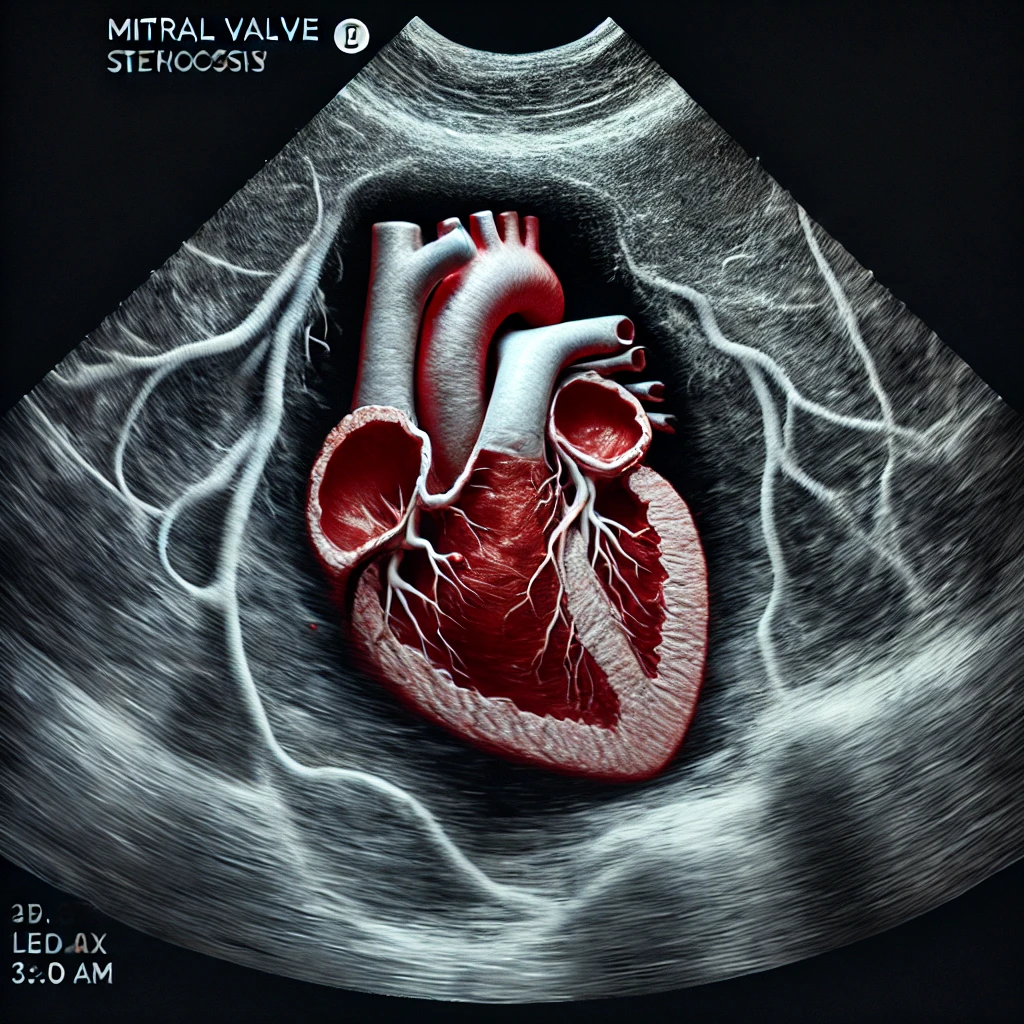
Mitral stenosis usually results from rheumatic heart disease, leading to fibrotic changes, commissural fusion, leaflet thickening, and sometimes calcification. This anatomical alteration increases the pressure gradient across the mitral valve during diastole, resulting in increased left atrial pressure and potential pulmonary hypertension.
1. Key Echocardiographic Modalities and Parameters
Echocardiography assesses both the structural and functional aspects of the mitral valve and associated hemodynamic changes. The primary modalities include:
- 2D Transthoracic Echocardiography (TTE)
- 3D Echocardiography (for anatomical detail)
- Doppler Echocardiography (for flow and pressure gradients)
- Transesophageal Echocardiography (TEE) (for detailed assessment, particularly pre-surgical evaluation)
2. Echocardiographic Parameters in Mitral Stenosis
| Parameter | Method | Normal Range | MS Severity Indicators |
|---|---|---|---|
| Mitral Valve Area (MVA) | Planimetry, Pressure Half-Time, Continuity Equation | >4 cm² | Mild: 1.5-2 cm², Moderate: 1-1.5 cm², Severe: <1 cm² |
| Mean Gradient | Continuous-Wave Doppler | <5 mmHg | Mild: <5 mmHg, Moderate: 5-10 mmHg, Severe: >10 mmHg |
| Pressure Half-Time (PHT) | Doppler (Deceleration Slope of E-wave) | 30-60 ms | Mild: 90-150 ms, Moderate: 150-219 ms, Severe: >220 ms |
| Pulmonary Artery Systolic Pressure (PASP) | Tricuspid regurgitation velocity + RAP | <30 mmHg | Mild: <30 mmHg, Moderate: 30-50 mmHg, Severe: >50 mmHg |
| Left Atrial Size | 2D Measurement (LA volume indexed to BSA) | 22-34 mL/m² | Enlarged LA suggests chronic increased pressure |
3. Step-by-Step Assessment Protocol
A. Mitral Valve Area (MVA)
- Planimetry: In a parasternal short-axis view, the mitral orifice is traced at the leaflet tips during diastole. 3D echo provides accurate planimetry, especially in irregular orifice shapes.
- Pressure Half-Time (PHT): The PHT is derived from the deceleration slope of the E-wave in Doppler imaging.
- Formula: MVA=220/PHT
- Continuity Equation: Utilized in cases with concomitant aortic valve disease; measures the stroke volume through both mitral and aortic valves.
B. Mean Pressure Gradient
- Doppler Gradient: Obtained from the continuous-wave Doppler across the mitral valve in an apical 4-chamber view. Gradients >10 mmHg indicate severe MS.
C. Pulmonary Artery Systolic Pressure (PASP)
- Elevated PASP correlates with severe MS and increased left atrial pressure. Estimated from tricuspid regurgitation velocity (TRV) and adding right atrial pressure (RAP).
D. Left Atrial (LA) Size
- LA dilation is a compensatory response to chronic pressure overload. It is measured in the parasternal long-axis view and quantified as LA volume indexed to body surface area (BSA).
4. WILKIN SCORE
The Wilkins Score (also known as the Echocardiographic Mitral Valve Score) is a scoring system used to evaluate the suitability of a patient with mitral stenosis for Percutaneous Balloon Mitral Valvotomy (PBMV). This score assesses the mitral valve anatomy based on four echocardiographic features, assigning points from 1 to 4 for each feature. Lower scores indicate favorable valve anatomy for PBMV, while higher scores suggest limited success and higher risk of complications.
Wilkins Score Components
- Leaflet Mobility
- Leaflet Thickening
- Leaflet Calcification
- Subvalvular Thickening
Each feature is graded on a scale from 1 to 4, where:
- 1: Mild
- 2: Mild-to-moderate
- 3: Moderate-to-severe
- 4: Severe
Detailed Scoring Criteria
| Feature | Grade 1 | Grade 2 | Grade 3 | Grade 4 |
|---|---|---|---|---|
| Leaflet Mobility | Highly mobile with minimal restriction | Mobile but limited at leaflet tips | Minimal mobility, most of leaflet affected | No or minimal forward movement |
| Leaflet Thickening | Leaflets near normal thickness (≤5 mm) | Thickening at mid-portion (5-8 mm) | Thickening at entire leaflet (8-10 mm) | Marked thickening (>10 mm) |
| Leaflet Calcification | No calcification | Small calcific areas in leaflets | Calcification noted throughout leaflets | Extensive calcification throughout |
| Subvalvular Thickening | Minimal chordal thickening | Thickening extending up to 1/3 of chordal length | Thickening extending up to distal 2/3 | Extensive thickening throughout chordae |
Total Score Interpretation
The scores for each feature are added to give a total score ranging from 4 to 16.
- Scores ≤8: Good candidate for PBMV, high likelihood of success.
- Scores 9-11: Intermediate candidates; PBMV may still be possible but with moderate risk.
- Scores >11: Poor candidate for PBMV due to high risk of complications and limited improvement.
Clinical Implications
- Lower Wilkins Scores indicate pliable, less calcified valves with minimal subvalvular thickening, making them ideal for balloon valvotomy.
- Higher Wilkins Scores often suggest significant leaflet immobility, thickening, or extensive calcification and subvalvular disease, generally making surgery a more favorable option.
5. Additional Parameters for Comprehensive Assessment
| Additional Feature | Relevance in MS | Assessment Technique |
|---|---|---|
| Commissural Fusion | Reflects rheumatic involvement | 2D & 3D echocardiography |
| Subvalvular Apparatus Thickening | Indicates disease chronicity | 2D echocardiography |
| Mitral Annular Calcification (MAC) | May complicate surgical repair | Parasternal long-axis view |
| 3D Echocardiography | Accurate MVA and leaflet morphology | Multiplanar reconstruction |
| Color Doppler | Visualize turbulent flow across mitral valve | Apical views |
6. Grading Severity of Mitral Stenosis
| Severity | Mitral Valve Area (MVA) | Mean Gradient (mmHg) | Pressure Half-Time (PHT) | PASP (mmHg) |
|---|---|---|---|---|
| Mild | 1.5-2.0 cm² | <5 mmHg | 90-150 ms | <30 mmHg |
| Moderate | 1.0-1.5 cm² | 5-10 mmHg | 150-219 ms | 30-50 mmHg |
| Severe | <1.0 cm² | >10 mmHg | >220 ms | >50 mmHg |
7. Role of Transesophageal Echocardiography (TEE)
TEE is valuable in detailed assessment, particularly for:
- Thrombus Detection: Especially in the left atrial appendage in atrial fibrillation.
- Better Visualization of Valve Anatomy: Allows precise assessment of leaflet motion, fusion points, and subvalvular structures.
- Pre-Procedural Planning for Percutaneous Balloon Mitral Valvotomy (PBMV): Essential to assess suitability and exclude LA thrombus.
Key Roles of TEE in Mitral Stenosis
- Left Atrial Thrombus Detection:
- TEE is highly sensitive for detecting left atrial thrombus, especially in the left atrial appendage, which is a contraindication for PBMV.
- Mitral Valve Anatomy Assessment:
- Provides superior visualization of the mitral leaflets, subvalvular apparatus, and commissures. Helps assess leaflet mobility, thickening, and calcification more precisely.
- Useful for scoring valve morphology (Wilkins Score) to determine PBMV suitability.
- Subvalvular Apparatus Evaluation:
- Offers detailed images of chordal thickening and fusion, which can impact procedural outcomes if severe.
- Mitral Valve Area (MVA) Calculation:
- Planimetry on TEE can provide accurate MVA measurements, especially in irregular or small mitral orifices.
- Other Assessments:
- TEE can help confirm the severity of MS by assessing flow velocities and gradients.
- Useful for evaluating associated mitral regurgitation (MR) or concurrent aortic valve pathology.
Indications for TEE in MS Assessment
- Inconclusive or poor TTE images.
- Suspected left atrial thrombus prior to PBMV.
- Preoperative or pre-interventional planning in severe MS cases.
- Detailed assessment of complex valve anatomy when surgical intervention is considered.
