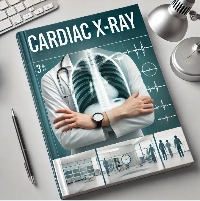
CARDIAC X RAY

About Course
Cardiac X-Ray Interpretation
The presentation provides an in-depth guide on interpreting chest X-rays (CXR) for heart-related conditions. It covers both technical and clinical aspects of X-ray imaging in cardiology.
Key Sections:
- Basics of CXR:
- Technical Quality (RIPE Criteria):
- Rotation: Ensure medial clavicles are equidistant from the spine.
- Inspiration: Adequate effort shows six anterior or ten posterior ribs.
- Projection: PA (preferred) vs. AP view impacts visibility and heart size.
- Exposure: Vertebral bodies should barely be visible.
- Technical Quality (RIPE Criteria):
- Structures Identified:
- Anterior View: Includes heart chambers, pulmonary vasculature, and aorta.
- Lateral View: Highlights the retrosternal space, IVC, and pulmonary veins.
- Heart Size and Cardiac Situs:
- Cardiothoracic Ratio: Normal is up to 50% in adults.
- Situs: Identifying organ positions for congenital anomalies.
- Chamber Enlargement:
- Right Atrial Enlargement: Vertical height >50% of the right heart border.
- Left Atrial Enlargement: Signs include double atrial shadow and carinal widening.
- Ventricular Enlargement: Distinct patterns for left and right ventricle enlargement.
- Pulmonary Vasculature:
- Features like venous/arterial hypertension, vascularity changes, and plethoras/oligemia.
- Cardiac Calcification:
- Differentiating pericardial vs. myocardial calcifications and identifying prosthetic heart valves.
- Congenital Heart Diseases:
- ASD, VSD, PDA, TOF, TAPVC, TGA: Characteristic CXR findings like “Egg on a String” for TGA and “Snowman Sign” for TAPVC.
- Other Pathologies:
- Pericardial Effusion: “Water bottle” appearance and narrow vascular pedicle.
- Valvular Diseases: CXR changes in mitral and aortic valve diseases.
- Special Equipment:
- Identifying pacemakers, ICDs, and prosthetic valves on X-ray.
- Clinical Case Examples:
- Real-world CXR images for conditions like pericardial effusion, situs inversus, and cyanotic heart diseases.
Course Content
CARDIAC X RAY
-
00:00
-
00:00
Earn a certificate
Add this certificate to your resume to demonstrate your skills & increase your chances of getting noticed.

Student Ratings & Reviews

No Review Yet